|
|
 |
 |
|
In technical collaboration with the Fyodorov Eye Microsurgery Institute,
Moscow,
The Sangubashi Eye Clinic, Shimouma, Tokyo, has developed Lasik Flapless (
corneal abrasion is made by a broad beam laser, and does not use any knife or
surgical instrument ) myopic refractive surgery using only an excimer laser.
The Fyodorov Institute has conducted more than 100,000 cases of PRK treatment
since 1987.
In brief, Lasik Flapless is myopic
refractive surgery, by flattening the convex curve at the center of the cornea
through abrasion of an optical zone of about 6.7 mm in diameter with a volume
three dimensional excimer laser.
Due to using Lasik Flapless, we reached a high accuracy rate in refractive
effect of more than 93% and get as close as possible to 100% using regenerative
tissue control during post operative care.
It is highly safe and effective.
Depending on the degree of myopia and
corneal thickness, the depth and the area of abrasion vary for different
patients.
In cases of extreme myopia and very thin cornea, Lasik Flapless re-operation
may be called for.
|
The Process of Lasik Flapless
|
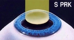 |
A picture showing the broad beam abrasion of the cornea surface.
The broad beam Excimer laser creates a smooth corneal surface.
The patient only needs to look at the
light for about 30 seconds and its finished. |
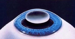 |
The post operative corneal surface which will recover within a couple of
days, is protected under a contact lens. |
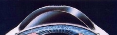 |
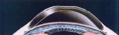
The flattened curvature of the cornea. |
|
@
|
| The
Process of Lasik |
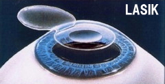 |
First of all make a flap from the cornea by microkeratome. |
 |
After the corneal flap is opened, and the cornea is exposed it is abrased
by a tiny scanning laser.
The corneal flap is returned to it's original position. |
 |
This shows the flattened curvature of the cornea post Lasik. |
|
|
Lasik Flapless VS Lasik, Video Presents 
|
 |
|
|
