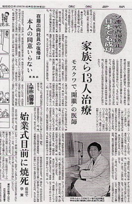
 ICO第26回シンガポール国際眼科学会 シンガポール RK後7年の角膜内皮細胞
ENDOTHELIAL CELL IN ARK PRACTICE
UP TO 7 YEARS
DIRECTOR M.D.
KODO OKUYAMA
SANGUBASHI EYE CLINIC GOTANDA
Institute of Refractive Keratoplasty
Summary: In l983 I Iecived Anterior Radial Keratotomy to both eyes at the moscow Research Institute of Eye Mierosurgery from Professor S.N. Fyodorov. 1)
I want to report on the excellent results achieved over the past 7 years using ARK on patients in Japan. Also I would like to show the complete safty of this procedure. Using a speculer microscope and putting the data through corneal endothelial cell computer norphologic analyzer.
I will present the histories of 17 eyes in 9 patients. One eye was uemotropic and didn’t need correction. These 9 patients were all members of my family or my friend’s families. All these patients gave informed consent.
method : ARK was performed on l7 eyes of 9 patients. The ages of all the subjects at the time of completion of this study are over 25 years old (average 32.5 years) and all of them had more then -2.50 diopters myopia.
Three out of the nine wore contact lenses regulary. (2RL,5RL and 9RL)
All cases except 3R had no previous history of disease. Case 3R had keratitis, while wearing a hard contact lense at the age of 22. All of the patients had naked vision less than 0.3 and a corrected visual acuity of more than1.0. Before ARK a video and written matter were used to explain the procedure to the patients. All patients are given corneometry as well as the standard examinations. The standard procedure developed by Prof. Fyodorov4) usd for ARK seven years ago and photographs were obtained of corneal endothelial cells using a Konan specular microscope and a morphometric cell analyzer, used to obtain cell counted the degree of polimegethism and pleomrphism. Case 2R,5RL and 9R were reoperatet and one case 9L was reoperated twice with the purpose of increasing the effect.
The magnification is calibrated with a micrometer scale. Thirty cells are outlined. The cells are digitized by touching the cell apices with a graphic tablet pen. These coordinates are entered on to a digitizer table and analyzed by computer for cell density, standard deviation (SD), coefficient variation (CV), average of cell area (AVE), maximum of cell area (MAX), minimum of cell area (MIN), and a histogram is made. .
Cell density is calculated by dividing one milion by the mean cell area.
CV is calculated by dividing the SD of cell area by the mean cell area.
Polimegethism is assessed indipendently of cell size using a dimentionlless index for CV. ‘Normal endothelial cells are about 300 microns square in size and hexagonal. Inflamation or injury can reduce cell count, hexagonality and uniformity of size. Eyes with exessive deviation in any of the above are not considered as candidates for ARK.
Result: Visual acuity in all eyes having had ARK had stabilized after four months. (Fig.2,3 and4) Tables 3 and 4show change in corneal refraction and visual acuity before and 7 years after. According to the size of the optical zone and the number of cuts, the resulting corneal refractive power can be decreased from 1 to 8 diopters. Out of 17 cases, one case (6L) of superficial keratotitis occured. It was treated with a two week course of gentamycin subconjunctive injection and a wide-spectrum antibiotic. After 7 years there are no complications and the cornea remains transparent. (Fig. 5)
Specular microscopic analysis shows mean cell densities of 2,347 cells per square mm between the incisions, standard deviation 122, coefficient variation 3l, average of cell area 426, maxmum of cell area 658, minimum of cell area l73 microns square. (Fig.6)
Slight increases in nonhexagonal cell (pleomorphism), and a variation in cell size (polimorphism) are observed. Case 3R had a history of keratitis.(Fig.7)
Specular microscopic analysis shows mean cell densities of 1,869 cells per square mm between the incisions, standard deviation 151, coefficient variation 28, average of cell 635, maxmum of cell area 818, minimum of cell area 162 microns square.(Fig.8) As the specular microscope only became available in 1986, preoperative data is not available for l7 cases, including this case. We can keep record of yearly rates of cell loss.
For 121 eyes in 61 patients from l987 to l988 the immediate cell loss a result of operating is 5.9%.
Discussion : Since the first pioneering work done by Professor Fyodorov and others in 1974, over 327,000 people have recived ARK.3) There have been, however, only limited reports of long term effects and sporadic reports on safty and effectiveness of ARK. Because of high satisfaction and lack of complications and the subsequent non-return of patients to the clinic, ther.e is limited statistical information available. There is in the case of Japan a certain resistance in the old establishment to ARK, because of the unfortunate experience with Professor Sato’s posterior radial ke.atotomy (PRK) in 1936. Therefore I make this report.
The result of ARK remains constant between 4 months and 7 years after operating. The changing refraction is limited to 8 diopters. The patient must be informed of this prior to operating. In six cases with previous
mild myopia aged 35 to 40 years, none wore glasses or contact lenses after seven years. (3RL,4RL,5L,6R,7L and 8RL) .
In previously moderate or severe cases of myopia, low grade glasses are worn when driving, at theatre or in some cases constantly, which doesn’t affect the field of vision as those wore previously. So nine cases discontinued use of contact lenses after ARK. Those that required lenses, were first examined at eight to twelve months with a specular microscope as a precautionar.y check.
Sometimes temporary glare and or starburst effect are noted.
Post-operative astigmatism not requiring corrective lenses was noted in cases 3R, 4R and 5RL. In cases 3R and 4R are 0.75 diopters, 5R is 1.00 diopter and in 5L is 1.50 diopters.
I’d like to make one final point about endothelial cell dynamics over the long term after ARE. Bullous keratopathy is not a possible result postoperatie endothelial cell loss of 5.9% , thereafter continuing at a
normal rate coinciding with Dr.Murphy’s 0.35 to 0.71% per year.(Fig.7)
Also cases reoperated showed a similar cell loss. At this rate of endothelial cell loss, a person would have to live to the age of 166 years before the critical level of 500 cells per square mm would be reached.
So specular microscopic examination is done before operation in order to facilitate obtaining informed consent as well as obtaining data to follow up post-operative cell loss and also the yearly rate of cell loss.
In the case of finding unusualy high levels of polymegethisn or polimozphism, these people are rejected for ARK. We find as did Dr.Scott, a higher incidence of such conditions among long term contact lens wearers. (Fig.6)
This could be a result of previous histories of infection.
We mesured endothelial cell loss and plotted the information for 121 eyes in 61 patients on a graph similar to Dr.Myer’s endothelial cell depletion graph.(Fig.2) On his graph a 35 year old person will have 2700 endothelial cells per square mm. If such a person loses 5.9% as an immediate result of ARK and we calculate natural aging cell loss of 0.35 to 0.71% ( according to Dr. Murphy ), then at the age of forty-two he should have 2,457 endothelial cells per square mm. But in practice our forty-two year old patient has 2,816 endothelial cells per square mm in his right eyes and 3,355 cells in his left eye.(Fig.9,10) In the other seven cases we recorded a similar high result. (Fig.11) One case 3R was lower and should continue to be observed over the long term.
Up till now 1,500 patients have received ARK at our clinic. (Fig.l2)
References :
i) K.Okuyama : myopia is possible to operate within 15 minutes.
Tokyo, Kobunsha p92-150, 1985
2) D.J.Mayer : Clinical wide-field specular microscopy.
Bailliere Tindall, London p52-53, 1984
3) S.N.Fyodorov, A.I.Ivashina : Microsurgery of the eye- Main aspects.
Mir. Publihers. Moscow p68-70, 1987
4) S.N.Fyodorov, V.V.Durnev : Operation of dorsaged dissection of corneal
circular ligament in cases of myopia of mild degree.
Annals of Ophthalmology p1986-1989, l979 Dec.
5) T.Yamaguchi et al. : Bullous keratopathy after anterior-posterior radial
keratotomy for myopia and myopic astigmatism.
Am. J. Opthalmol. 93:600. 1982
6) Scott M Mac Rae et al. : The effects of hard and soft contact lenses
on the corneal endothelium.
Am. J. Ophthalmol. p50-57, July 1986
7) Scott M. Mac Rae et al. : The effect of radial keratotomy on the corneal
endothelium.
Am. J. Ophthalmol. p538-542, October, l985
 ICO第26回シンガポール国際眼科学会 シンガポール RK後7年の角膜内皮細胞
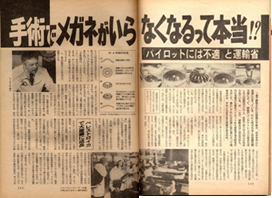
 消費者問題ジャーナリストの船瀬俊介さん(39)は、このRK(Radial Keratotomy)手術を今年2月末から4月末にかけて右目、左目の順に片方ずつ受けた。左目の手術からもすで2カ月以上たつが、「いま、気分はオートフォーカス。遠くも近くもクッキリ見えて最高です」と喜んでいる。かつて0.04だった右目は、いまや1.5になり、0.06だった左目が1.0にまで回復したのだから無理もない。なにしろ、以前は視力検査表の一番上の大きな文字か何歩も前進しなけれは読めず、大学時代から分厚いレンズの眼鏡を手放せなかった。
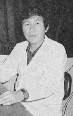 「手術で近視が治る」という話は以前、耳にしたことがあるし、「下町の赤ひげ」と呼はれていた奥山医師の父とも親しかった船瀬さんだが、最初はやはり半信半疑で、特に、「角膜を切る手術だと聞いてからは怖くて、返事をのらりくらりと引き延ばしていました」という。しかし、消費者運動の活動家であり、好奇心旺盛な船瀬さんは、その後も手術の安全性や効果について、奥山医師らに積極的に“取材”。「すでにアメリカで30万例、ソ連で12万例ほど行われていて、重大な後遺症はゼロ、失明例は麻酔のやりすぎによる1例だけで、これも手術以前の問題だし・・・・、それに、自分自身この手術を受けて近視を治しておられる奥山先生の人柄を信頼したんです」と、今年初め決心したそうだ。
 それが1960年代末、ソ連の眼科医、S・N・フィヨドロフ博士らによる研究、開発で、内皮細胞を傷つけないよう角膜の前面だけに切り込みを施すという、より弊害の少ない新しい術式として甦ったのだ。
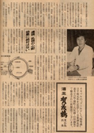 アメリカでは、1978年、学界の専門誌でこの術式を知ったデトロイトのL・ポアーズ医師ら、眼科の開業医たちがとびつき、それぞれの医師が改良を加えながら、ソ連を凌ぐ勢いで広まっている。
 聖路加国際病院の山口達夫・眼科副医長は、この手術が米国で爆発的に広まったとき、サルを使った実験で、角膜表面だけを切っても内皮細胞がやはり一定程度は滅少することを発見、早くから警告していた。
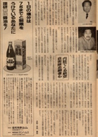 こうした指摘に対して奥山医師らRK推進の医師らは、「視力の不安定さはふつう3カ月、グレアは良い人でも10カ月で、落ち着き、なくなる。外傷に対する強さも、RKを二回受けたパイロットが激しい事故で顔に衝撃を受けても、目だけは大丈夫だったという報告まである」と、データを挙げて反論。

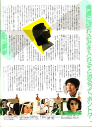 手術で近眼治るなんて・・・ウソみたいな話である。上智大学外国語学部三年生の内藤るりかさん(二十二歳)も初めてその話を父親から聞かされたときは、半信半疑だった。
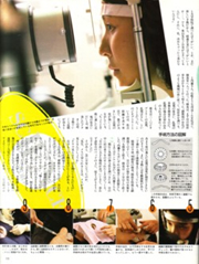 心配そうな河辺くんを尻目に、検査室へ入って行った。検査は、まず目の屈折率をはかることから始まった。顕微鏡のお化けのような機械の前に座って、屈折率を調べる。近視の度合、乱視の有無を検査するのだ。乱視の場合は切れ込みの入れ方が変わってくる。
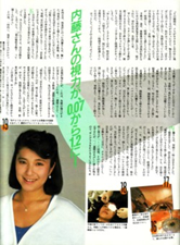 内藤さんの手術は一週間後の五月二十二日(水)に決定。ところが、その間に、とある週刊誌に近眼矯正手術の後遺症で失明者が続出、という記事が掲載された。
 1985年5月12日発行 サンデー毎日
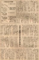 強度の近視に悩んでいた奥山さん、その情報に飛びついた。そして調べてみると、それは、モスクワ眼科マイクロサージュリ研究所のスヴイエットスラフ・ニコラエビッチ・フョードロフ教授が、実施しはじめたばかりの手術だということがわかった。
 したがって、入院する必要などは全くなく、通院でかまわない。手術当日だけは眼帯をしていなければならないが、翌日はとりはずせる。_というほど簡単なものだ。